Friday, May 30, 2008
Wednesday, April 16, 2008
M.I. ADMINISTRATIVE PROFESSIONALS
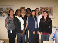
Many thanks to the group of volunteers from the Joint Department of Medical Imaging who have used their creativity and worked hard in preparing for this week to celebrate our Administrative Professionals!
ADMINISTRATIVE ASSISTANTS AND SECRETARIES
 Administrative assistants and secretaries are a great asset to the Medical Imaging Department. They support radiologists, charge technologists, supervisors and managers. Their daily responsibilities play an important role in ensuring that standards of organization and quality are met and surpassed consistently.


The patient flow coordinators are responsible for the overall organization of a patient's visit to the Medical Imaging Department. They coordinate and facilitate the efficient flow of patients throughout the various stages of their visit. They also act as a liaison between the patient, technologist, nurse and radiologist.
The appointment schedulers are responsible for booking appointments for all modalities across multiple sites. This group is efficient, able to cope with high volumes of calls, and accurately inform patients on preparation for their exams. They also play a key role in communicating information to the radiologists, technologists and nursing staff.

 MEDICAL IMAGING BILLINGS, CALL CENTRE AND SUPPORT SERVICES

 As integral members of the Department of Medical Imaging, the billing clerks work diligently to keep information flowing to the Ministry of Health so that financial responsibilities are captured and met. This is crucial to our hospitals as this enables them to meet and exceed the standards of providing outstanding patient care. Although this group works behind the scene, they are invaluable assets to the department.
The Medical Imaging Call Centre acts as a liaison between referring physicians and medical imaging staff. They are responsible for locating radiologists, and facilitating the arrangement of urgent bookings. The main objective of the call centre is to improve overall communication and to continue the departmental goal of providing exceptional patient centered care.
The Support Services personnel are responsible for managing the release of patient information in medical imaging. This includes directing patient images, reports, and verbal results to the appropriate health care provider ensuring continuity of care. They are also responsible for the processing, sorting and retrieval of hard copy films.
SUPPORT SERVICES SUPERVISORS
The supervisors oversee the clerical and administrative staff in all Medical Imaging modalities. They participate in the development, implementation and ongoing maintenance of departmental policies, procedures and program initiatives. In their leadership roles, they strive to ensure that the goals and objectives of the department are met, focusing on a patient centered care approach and excellent customer service.
We hope this brief overview of the roles and responsibilities of these wonderful staff members has given you a better understanding of their importance within our joint departments. Please join us in helping them celebrate Administrative Professional's Week.
Sunday, March 04, 2007
PPL Corner
Julie can be found in room 3-970 at PMH, by phone at 16-4750, or by e-mail PPLMedicalImaging@uhn.on.ca. Check out the Professional Practice section of our department web site.

Wednesday, January 24, 2007
CT Scan
 Tomography comes from the Greek tomos (slice) and graphia (describing).
What do The Beatles and CT have in common?

EMI recording studio developed the first CT scanner.
The idea of CT Scan, computed tomography, or CAT scan as some people like to say, has been around since 1967. Alan Cormack of S. Africa developed the theory behind CT. Godfrey Hounsfield of EMI in England developed the first scanner. These two scientists shared the Nobel Prize in Medicine in 1979 for their discovery.
The 1971 prototype from EMI took 5 minutes to scan each image and over 2.5 hours to reconstruct the image data.
The first commercial EMI scanner was used to image the brain in 1972 in Wimbledon, England. It took about 4 minutes to scan the brain and 7 minutes to reconstruct each image. And we complain when patients won't hold still now for 12 seconds!

CT uses X-rays to obtain images and a powerful computer to reconstruct and manipulate these images. The patient lies on the scanner table and moves horizontally into the gantry. Inside the gantry is the x-ray tube and detector array. This x-ray tube and detector rotate inside the gantry as the patient slides through on the table; we can scan any body part from head to toe. A routine brain takes approximately 12 seconds to scan. A routine chest takes about 10 seconds to scan. An entire chest, abdomen and pelvis can be scanned in approximately 18 seconds. It only takes a few seconds to reconstruct the image data.
Some more advanced uses for CT Scan include CT Angiography, CT Guided Biopsy, CT Guided Radio Frequency Ablation (RFA), and Virtual Colonography. Some of these procedures are performed by a Radiologist. Most are performed by a Medical Radiation Technologist (MRT) under the supervision of the Radiologist. The scanner views your body like a loaf of bread. We can view axial images (like a cross-ways slice); sagittal images (like a lengthwise slice); or coronal images (like slicing the crust off). We can get really fancy when we do multiplanar reformats (MPR) or 3D imaging.
At MSH, there are 3 64 slice CT Scanners used in everyday operation. There is also 1 4 slice CT Scanner used for the Lung Screening Program, and as you read this entry, the 16 slice CT Scanner is scanning away in the Emergency department, operated by the Gen Rad team.
At PMH, there are 3 64 slice scanners. Two scanners run from 0800-2300hrs, Monday to Friday. The Gen Rad technologists are trained to scan Level 2 CT (single phase studies) and they are responsible for the daily operation of the other scanner. On an average, there are 80-90 patients scanned per day! For the most part, patients get the "PMH special": chest, abdomen, pelvis, head/neck with IV contrast-the works! This volume of patients would not be possible without one very special person- Stella, the CT assistant.

At TWH, the Medical Imaging department (specifically CT), the Neuroscience department as well as the Radiation medicine program work in conjunction to form Canada's first Gamma Knife centre. With the help of CT scans, beams of gamma radiation are targeted to areas of the brain where it's too dangerous or challenging to access.

There are 2 64 slice scanners on the 3rd floor operated by CT technologists and an 8 slice scanner in the Emergency department operated by General Radiography technologists. This is where the Regional Stroke Study centre relies on timely imaging of patients who might have had a stroke. If the stroke is detected in time, a special medicine is given to help in the recovery of patients.
At TGH, there are 3 64 slice scanners; one dedicated to Cardiac CT, where anatomy of the heart, coronary circulation and the great vessels are seen. The 3D lab team then will use the CT scan data to produce high resolution 3D images of the heart and great vessels like this one:


There is also a 4 slice CT scanner in the Emergency department that is operated by General Radiography technologists. There are a large number of research projects that are scanned at TGH as well as lung biopsies and pre/post transplant procedures.
The New Women's College Hospital is the newest addition to the corporate Medical Imaging family. Just last week, they said goodbye to their 4 slice scanner to welcome a new 64 slice scanner.
So, from 2.5 hours to 12 seconds, CT has come along way. On behalf of MSH/UHN/WCH's CT scan departments, we hope you enjoyed this brief overview of such a dynamic and technologically advanced section of healthcare!
Tuesday, September 12, 2006
Welcome Post
In the coming days and weeks we will begin posting messages, so check back frequently for updates.


